06 Apr

A temporally consistent atlas of neonatal brain development

Excited to present my work on temporally consistent modelling of the neonatal brain morphology from structural brain magnetic resonance (MR) images at the Centre for the Developing Brain at King’s College London, St’ Thomas Hospital.

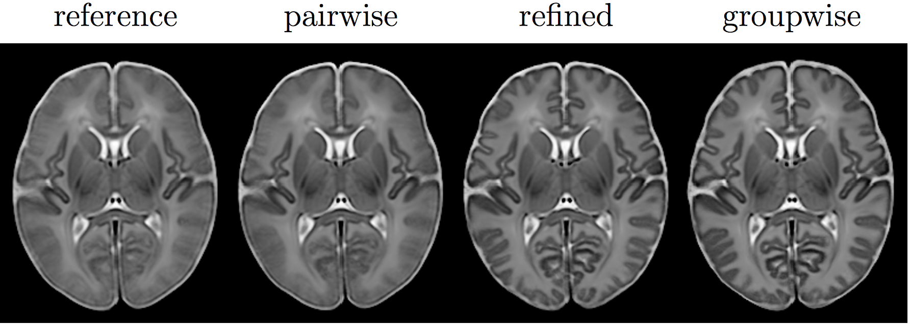
Axial mid-sections of mean intensity and shape templates at 40 weeks PMA.

After providing some background on brain atlas construction, the talk focuses on our novel group-wise approach for the construction of an unbiased spatio-temporal brain with improved temporal consistency, lower computational cost, and considerably higher cortical detail than previous neonatal atlas construction techniques. It is a summary of our work detailed in Schuh et al., “Unbiased construction of a temporally consistent morphological atlas of neonatal brain development”, preprint available on bioRxiv (doi:10.1101/251512).

11 Apr

dHCP: Neonatal Cortex Reconstruction

A preprint of our article “The Developing Human Connectome Project: a Minimal Processing Pipeline for Neonatal Cortical Surface Reconstruction”, covering the full structural image processing steps, following the MR volume reconstruction, from whole brain segmentation to the reconstruction and spherical mapping of the neonatal cortex, is now available on bioRxiv (doi:10.1101/125526).

10 Jan

Paper accepted at ISBI 2017

Our paper on deformable models for the reconstruction of the neonatal cortex from structural T2-weighted brain MR images was accepted for presentation at the 2017 IEEE 14th International Symposium on Biomedical Imaging (ISBI) which takes place this year April 18-21 in Melbourne, Australia.

A deformable model for the reconstruction of the neonatal cortex

Two stages of deformable mesh reconstruction of one hemisphere of a white matter surface. Colour depicts residual distance.

We present a method based on deformable meshes for the reconstruction of the cortical surfaces of the developing human brain at the neonatal period. It employs a brain segmentation for the reconstruction of an initial inner cortical surface mesh. Errors in the segmentation resulting from poor tissue contrast in neonatal MRI and partial volume effects are subsequently accounted for by a local edge-based refinement. We show that the obtained surface models define the cortical boundaries more accurately than the segmentation. The surface meshes are further guaranteed to not intersect and subdivide the brain volume into disjoint regions. The proposed method generates topologically correct surfaces which facilitate both a flattening and spherical mapping of the cortex.